Images from Science 2 Exhibition Opens at RIT on Oct. 11
Display features 61 images taken by scientists and photographers from various disciplines
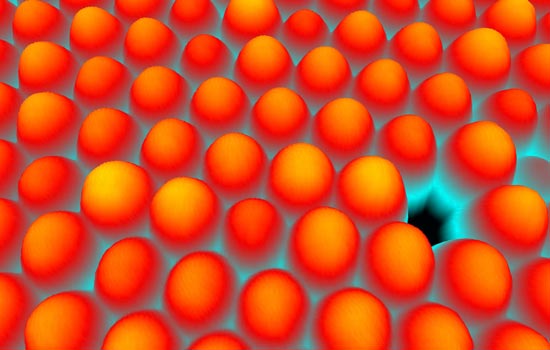
Hans U. Danzebrink, Atomic force microscope image
Dislocations in a photonic crystal arrangement of polystyrene nanospheres.
Acclaimed scientists and photographers from around the world will share their scientific research, discoveries and observations of natural wonders in an international photography exhibition opening next month at Rochester Institute of Technology.
RIT’s School of Photographic Arts and Sciences will host Images from Science 2 featuring 61 photographs from various scientific disciplines including astronomy, biology, engineering, medicine, oceanography, physics and nanotechnology. The opening, at 7 p.m. on Saturday, Oct. 11, takes place in SPAS Gallery, third floor of RIT’s Frank E. Gannett Building. The show runs through Oct. 25.
Viewers can see breathtaking images of the tiniest of things, from an unknown species of octopus measuring a half-inch in length to nanospheres with a diameter 300 times smaller than a human hair.
To accompany the display, RIT Cary Graphic Arts Press, the publishing arm of the Melbert B. Cary Jr. Graphic Arts Collection at RIT, produced a full-color companion catalog of all the images in the exhibition. The publication features an introduction by Martin Scott, a former director of scientific imaging at Eastman Kodak Co. The catalog will be available for purchase online through the RIT Cary Graphic Art Press at the RIT Press webstore and the Amazon webstore.
“The quality of the images is excellent,” says Michael Peres, RIT department chair of biomedical photographic communications and one of the exhibit organizers. “All the traditional imaging methods are utilized, including micrography, high-speed nature photography, macro photography, but also some obscure methods such as scanning tunneling microscopy and radiography. It’s fascinating to see what people are currently doing in their respective scientific fields and the types of images they are producing.”
Scientists, photographers and worldwide institutions submitted entries for consideration. A photograph from Lennart Nilsson, a pioneer in medical photography, is among the images in the new exhibit. The award-winning Swedish photographer and scientist has been a leading supporter of the project since its inception. An international selection committee chose the 61 images from more than 300 entries. The final images were selected based on their scientific content, aesthetics and difficulty in making of the photograph.
In fall 2002, RIT launched the inaugural Images from Science exhibition. Since that time, Images from Science, has been hosted by 23 organizations in seven different countries, most recently in the Czech Republic.
“The first exhibition was so successful and far reaching because of the work produced by its outstanding contributors,” says Andrew Davidhazy, RIT department chair of imaging and photographic technology and one of the exhibit organizers. “Its longevity can be attributed to the stunning photographs that depict life as it is seldom seen by the general public. With this second exhibit, we wanted to once again emphasize to the photographic community that images made other than for artistic purposes can be appreciated not only for their scientific content, but also for their aesthetics.”
Adobe Systems Inc., Carl Zeiss MicroImaging Inc. and Durst Image Technology are sponsoring the project.
For more information, call RIT’s School of Photographic Arts and Sciences at 585-475-2863 or visit the Images from Science Web site.
Credit and caption information for Images from Science 2 photographs:
Aesthetic Imperfections, 2008
Hans U. Danzebrink, Atomic force microscope image
Caption: Dislocations in a photonic crystal arrangement of polystyrene nanospheres
Octopus Paralarvae, 2000
David Paul, photograph, color positive film
Caption: Photographed on the Coral Sea, Far North Queensland, this octopus paralarvae is a pelagic juvenile of an unknown species collected at night over deep water. Actual size is about 15 mm top to bottom.
Recommended News
-
April 18, 2024
!['an arial view of Croatia’s Neretva Delta is shown through clouds with birds in the air.']()
RIT Global students set to present at Imagine RIT
Students from RIT’s global campuses will be making the trek to the U.S. for this year’s Imagine RIT: Creativity and Innovation Festival. Teams from all of RIT's global campuses will be presenting their exhibits in the Gordon Field House; this is the second year that students from global campuses will be attending the festival in person.
-
April 18, 2024
!['a headshot of Alireza Vahid appears on the right with his name, title, and department to the left.']()
Engineering professor becomes part of SMART Hub to improve wireless spectrum accessibility
The wireless spectrum has become very crowded real estate, and work is underway through a new technology research center to improve spectrum access, co-existence, and security.
-
April 17, 2024
![a generated image of the new main concourse of a staium is displayed with the field to the left and a concession stand to the right.]()
RIT to break ground on new athletic stadium
Rochester Business Journal talks about the new athletics stadium being built on the RIT campus.
-
April 17, 2024
![Three biologists in a wooded area gaze up toward the treetops, looking for birds.]()
Should We Change Species to Save Them?
The New York Times cites a paper co-authored by Evelyn Brister, professor in the Department of Philosophy, about Conservation science.


