RIT, URMC partner on cancer screening technology
RIT and UR collaboration shines light on photoacoustic imaging
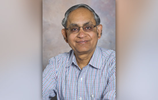
Navalgund Rao
A medical imaging technology that holds promise for screening prostate and thyroid cancers begins with a pulse of light.
Scientist Navalgund Rao, from Rochester Institute of Technology, has advanced the capability of photoacoustic imaging, a hybrid technique that combines light and ultrasound waves. His prototype imaging system focuses the waves through an acoustic lens that functions like an optical lens on a camera. The acoustic lens sharpens the contrast between normal and abnormal tissue for the early detection of certain cancers.
Rao, a professor in RIT’s Chester F. Carlson Center for Imaging Science, collaborates with Vikram Dogra, M.D., professor of radiology and urology in the University of Rochester Medical Center Department of Imaging Sciences. While Rao provided the technological solutions, Dogra focused on areas of need.
“In prostate imaging, ultrasound typically is used, but it doesn’t give a good contrast,” Rao says. “You can barely see a suspected lesion. And because you can barely see it, the only way to confirm it is to do a biopsy. And if you cannot see it properly, you can’t do a biopsy, and you have to go in and take many regions in the prostrate, called a multi-core biopsy.”
Ultrasound imaging reads the reflection of unfocused sound waves sent into the body; photoacoustic imaging pulses near-infrared light into the body to make ultrasound waves in the biological tissue that absorbs it. The tissue expands and contracts, creating acoustic pressure waves that propagate in all directions, Rao says.
“There was a need,” he says. “There is no other imaging modality that can be used in a screening mode. We initially thought of targeting prostate imaging with photoacoustic to see whether the cancer can be visualized better because the contrast comes from the absorption of the laser light in the near infrared and blood has a very high absorption co-efficient compared to the rest of the tissue. Tumors are very well saturated with blood so it was our hypothesis that they would stand out.”
The tunable laser in the prototype devices collects data along multiple wavelengths within the near-infrared spectrum. The optical absorption in the hemoglobin—the blood content—can be quantified at every pixel location to reveal distinct signatures of molecules within the blood, yielding a finer distinction between biological tissues rich in or depleted of oxygen.
Deoxygenated hemoglobin was “much higher” in the cancerous lesions than in the non-cancerous tissue samples Rao and Dogra imaged in a yearlong study funded by the Radiology Society of North America. The researchers used their patented imaging system to scan 50 cancerous and normal thyroid samples and 80 cancerous and normal prostate samples in the study conducted last year.
Rao and Dogra worked with pathologists at URMC to verify the accuracy of their study. They compared abnormal signals in their scans with the regions of tissue samples the pathologists marked as diseased.
“Tumors are very well saturated with blood so it was our hypothesis that they would stand out because cancer cells are hungry and take up oxygen very fast,” Rao said. “This study showed us that the deoxygenated content was much higher in the cancerous lesion compared to the normal, non-cancerous tissue. That was our significant finding. This large photoacoustic imaging study was probably the first one that has ever been done. No one else has done photoacoustic imaging this way.”
RIT and the UR hold a joint patent on Rao and Dogra’s device. The researchers created the start-up company Advanced Acoustic Imaging Technologies and are seeking funding to make their device ready for clinical testing. Working with Rao are research associates Chinni Bhergava, an RIT alumnus with a degree in electrical engineering, and Saugata Sinah, a Ph.D. student in RIT’s Chester F. Carlson Center for Imaging Science.
“This technology has potential uses for prostate and thyroid cancers; those are the two we have investigated,” Rao says. “It can also be used in breast imaging and skin imaging. There are a lot of possibilities and each one will require a different design.”
Another possibility involves injecting a targeted contrast agent, a chemical molecule that would attach itself to a specific cancer cell.
“And to the same molecule, if we can attach a photoacoustic agent, like a near infrared dye, then—during photoacoustic imaging—the agent will pinpoint the location and broadcast that it’s sitting on top of a cancer cell,” Rao says.
The project has been funded, in part, by grants from the Empire State Development’s Division of Science, Technology and Innovation (NYSTAR) and the Center for Advanced Technology, the Radiology Society of North America and an instrumentation grant from Lang Memorial Foundation, awarded through the RIT Chester F. Carlson Center for Imaging Science.
Rao currently lives in Brighton with his wife Lakshmi and his extended family.
