Imaging science student wins ‘best paper’
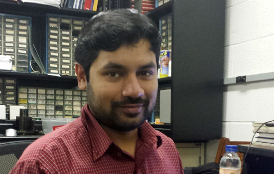
Graduate student Saugata Sinha won Best Student Paper at the 2013 IEEE Western New York Image Processing Workshop for his work on detecting thyroid cancer.
A novel technique for detecting thyroid cancer was the topic of the Best Student Paper award at the 2013 IEEE Western New York Image Processing Workshop held on Nov. 22 at Rochester Institute of Technology.
Graduate student Saugata Sinha, a Ph.D. student in RIT’s Chester F. Carlson Center for Imaging Science, won the award based on reviewers’ recommendations and the presentation of his paper, “Differentiation between malignant and normal human thyroid tissue using frequency analysis of multispectral photoacoustic images.”
Conference organizer Nathan Cahill, associate professor in RIT’s School of Mathematical Science, described Sinha’s research and presentation as “great examples of the high quality we strive to achieve in this workshop.”
Photoacoustic imaging is a hybrid imaging technology in which laser light is absorbed by soft tissue to generate ultrasound waves. The nascent technique may emerge as a new method for screening thyroid and prostate cancers.
Sinha, originally from Kolkata, India, expects to earn his Ph.D. in summer 2014. His award-winning paper represents one aspect of his thesis research, which he conducts under Navalgund Rao, professor in the Center for Imaging Science, and Dr. Vikram Dogra, professor of radiology and urology in the department of imaging sciences at the University of Rochester Medical Center.
“Our preliminary results have shown that it is possible to differentiate between malignant and normal thyroid tissue using the frequency analysis technique of photoacoustic images taken at multiple light wavelengths,” Sinha said.
Previously, the team analyzed two-dimensional photoacoustic images based on intensity values in order to compare hemoglobin concentration in cancerous tissue with normal tissue.
“In this research, I have gone one step further and investigated the frequency content of the raw photoacoustic data, which can be related to shape, size and distribution of different light absorbing tissue constituents,” Sinha said. “The additional features provided by the frequency analysis technique can be combined with the features already extracted using intensity value based analysis and this combined set of features can be used for noninvasive characterization of tissue abnormality, which is our ultimate aim.”
Topics
Recommended News
-
April 19, 2024
![an orange and white sign reading Scavenger Hunt with a QR code is shown on an easel sitting in a common area outside in the academic space on campus.]()
Scavenger Hunt for RIT Students planned at Imagine RIT
RIT spices up Imagine RIT with an exciting scavenger hunt exclusively for students. Participants can follow clues to discover selected exhibits, snap selfies with QR codes, and compete for a chance to win one of three $100 Tiger Bucks prizes.
-
April 19, 2024
![groups of people are seen gathering for the groundbreaking of the new Tigers stadium on the RIT campus.]()
Rochester Institute of Technology breaks ground on new Tiger Stadium
The Democrat and Chronicle speaks to Jacqueline Nicholson, executive director of Intercollegiate Athletics, along with alumni, coaches, and lacrosse players about the excitement of a new home for men's and women's soccer and lacrosse teams.
-
April 19, 2024
![9 people and RITs mascot Ritchie stand under the shade of a tent in a line with shovels in front of a mound of dirt marking the groundbreaking of the new stadium.]()
RIT breaks ground on new soccer, lacrosse stadium
WHAM-TV covers the groundbreaking of the new Tiger Stadium on the RIT campus.
-
April 19, 2024
![Christy Tyler is pictured in front of a brick background talking to a reporter at News8 Rochester]()
RIT and URMC partner to study microplastic’s impact on the environment and human health
WROC-TV talks to Christy Tyler, professor in the Thomas H. Gosnell School of Life Sciences, about how climate change will impact microplastics.


