Innovative image de-noising method helps diagnose schizophrenia
By Amy Mednick
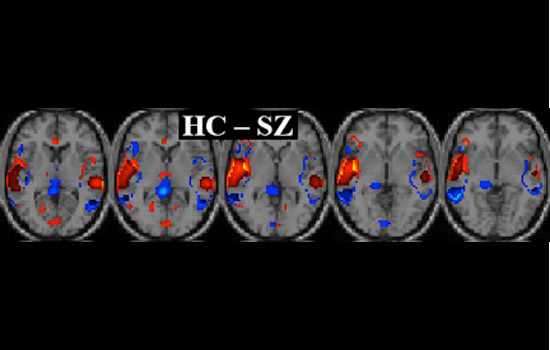
This series of images shows the difference between a healthy person’s brain versus a person with schizophrenia. The red areas on the brain template indicate the regions of a healthy brain involved in identifying and processing sounds while the blue areas indicate relatively more active regions of a schizophrenic brain. These images were obtained using Khullar’s image denoising technique in addition to other segmentation algorithms.
Clinicians across the world now have access to an image de-noising “toolbox” that will allow them to improve the diagnosis and treatment of patients with schizophrenia or bipolar disorder. This pre-processing algorithm enables scientists to compare, with greater specificity, brain scans of schizophrenic patients and healthy control subjects.
Siddharth Khullar, a second-year doctoral student at RIT’s Chester F. Carlson Center for Imaging Science, is pioneering new techniques that calibrate functional magnetic resonance imaging, commonly called fMRI, in precise detail, allowing scientists to discern brain functionality of schizophrenic patients better than ever before.
“Neurodiseases don’t show up in a brain scan as readily as, for example, a brain tumor,” says Khullar, who studies with Carlson Center Director Stefi Baum. “If someone has a symptom of schizophrenia, or a similar disease such as bipolar disorder, they can take a cognitive test inside the fMRI scanner and, with this de-noising method, clinicians are able to compare and analyze the resulting images more accurately with the same scan of a healthy person.”
Even as Khullar works with Baum at RIT, he lives in sunny Albuquerque, N.M. He works as graduate research associate in the Medical Image Analysis Lab led by Vince Calhoun at the Mind Research Network for Neurodiagnostic Discovery. Khullar received a master’s degree in electrical engineering at RIT in 2009, and then started the Imaging Science Ph.D. program and interned at the Mind Research Lab that summer. After a year of graduate classes at RIT, Khullar continued to work on his research at Mind Research Network and is currently funded by a federal grant from the National Institutes of Health.
During his first year at the Mind Research Network, Khullar has already published a journal article and presented his findings at three major medical imaging conferences. And, he says, another journal article is in the works.
“I am thrilled about our association with the Mind Research Network,” says Baum, who is also an astrophysicist. “These types of partnerships emphasize the interdisciplinary and collaborative nature of our work at the Center. The expertise that Khullar is developing as an imaging science student will help medical professionals’ understanding and diagnoses of schizophrenia and other mental disorders.”
The fMRI scan captures the level of blood flow in the brain over time, similar to capturing a movie of the brain. Ordinarily the image sequence produced is extremely difficult to quantify. Khullar’s method identifies and quantifies regions of activity in the fMRI brain images in a way that allows clinicians to differentiate characteristics of healthy and schizophrenic patients.
“We have shown, through our published work, that our algorithm is better in terms of preserving vital information about neural activation patterns within the brain,” Khullar says.
In his most recent research, Khullar is working on a new pre-processing methodology that allows clinicians to compare the patient’s brain activity at rest and while the patient is conducting a simple activity such as pressing a button in response to a noise.
Khullar’s new technique uses the observations of patient’s brain resting state activity to build an atlas representing the patient’s brain function that can be used to align the data obtained when the patient performs a task. This functional alignment enables improved fusion of data when studying a group of individuals who suffer from the same neurological ailment such as schizophrenia or even autism. The aim is then to use that fused data to better understand what is happening in the brains of individuals with specific neurological conditions.
“My inherent goal is to make a broader impact on mankind,” Khullar says.
Amy Mednick is a writer and editor based in Rochester, New York.
Topics
Recommended News
-
April 16, 2024
!['the interior of the Strong Museum is shown with people mingling on the left side of the frame and an informational table about the Rochetser School for the Deaf on the right.']()
Strong Museum hosts Deaf Day of Play
WHAM-TV talks to Danny Maffia, senior lecturer in NTID's Department of ASL and Interpreting Education and director of Interpreting, about the event.
-
April 16, 2024
!['Students from NTID are seen in the butterfly exhibit at The Strong Museum of Play.']()
Strong Museums’ Deaf Day of Play highlights inclusivity for those hard of hearing
Spectrum News covers the Deaf Day of Play at the Strong Museum of Play.
-
April 16, 2024
!['Joshua Sitte is shown in an auditorium standing for an interview.']()
Deaf Day of Play at Strong National Museum of Play
WHEC talks to Joshua Sitte, student interpreter at RIT, during the Deaf Day of Play at The Strong Museum of Play.
-
April 16, 2024
!['Three students stand in an open area of a university.']()
RIT’s Expressive Communication Center students explore ableism in communication-focused settings
RIT's Expressive Communication Center delves into the challenges of ableism in communication education, presenting their findings at a national conference in Arizona. ECC students aim to foster dialogue on accessibility and inclusive communication practices, advocating for more inclusive assessment methods in higher education.