Graduate Students
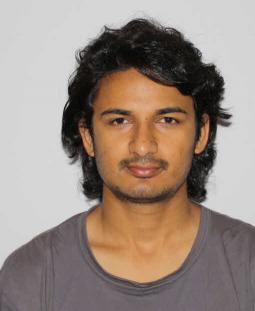
Shusil Dangi
PhD Candidate, Imaging Science (YR. 2)
Bio: Shusil is currently a PhD student in Imaging Science at Rochester Institute of Technology, NY, US. He recieved his Bachelor's Degree in Electronics and Communication Engineering (signal processing major) from Pulchowk Engineering Campus, Institute of Engineering, Kathmandu, Nepal. He was awarded the prestigeous "Prof. F.N. Trofimenkoff Academic Achievement Award" for his academic excellence throughout the Bachelor's degree. His undergraduate thesis was on "FPGA Implementation of Stereo Vision Based Unmanned Ground Vehicle".
Research Interests: Medical Imaging, Computer Vision, Image Segmentation, Multimodal/Monomodal Image Registration
Current project: Segmentation of the Left Ventrical (LV) from Tri-plane Time Series TEE data and LV volume reconstruction. The deformation field will be used to track LV features to fuse US and MRI images to provide better visualization during Intra-operative image-guided procedures.
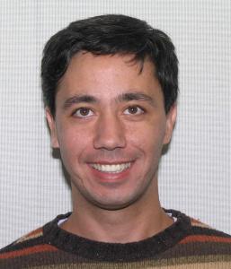
Kfir Ben-Zikri
PhD Candidate, Imaging Science (YR. 3)
(Summer intern co-advised with Dr. Nathan Cahill)
Bio: Kfir Ben-Zikri is research assistance as a PhD student at RIT in imaging science. He has his master in Electrical Engineering also from RIT, and his bachelor in Biomedical Engineering majoring in signal processing at Ben-Gurion University in Israel. He has work experience of four years as group leader of QA engineers in AMDOCS, working on the billing software of telecommunication companies, such as AT&T, Comcast, etc. He is married with twin’s girls 4.5 years old.
Research Interests: Medical Imaging, Algorithms Design, Prototyping & Implementation, Signal (Image) Processing, ill-posed inverse problems, Compress Sensing, Computer Vision, Computation Optics & Photography, Machine Learning, Information Theory, Bio-Sensors, 3D and Human Perception, Team work and Multidisciplinary Collaborations.
Current projects:
1. NIH grant supervised by Dr. Nathan Cahill, Dr. Maria Helguera (RIT), together with Kitware and Dr. Marc Niethammer (UNC). Apply geometric metamorphosis to longitudinal chest CT. Computational method based on image registration to determine pathology changes while compensating for non-rigid background deformations.
2. Segmentation of the Left Vertical (LV) from Tri-plane Time Series TEE Data and volume reconstruction for getting the deformation field of the LV. The deformation field will be used for tracking the LV and fusion with MRI during Intra-operative Image Guidance procedure.
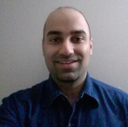
Aditya Daryanani
Masters Student, Computer Engineering (YR. 2)
Bio: Aditya is an MS student from the Computer Engineering department who is interested in Biomedical imaging and is investigating how computer engineering techniques such as computer vision, machine learning can be applied to help medical professionals make better decisions and save lives.
Research Interests: Machine Learning, Object Tracking and Segmentation, MRI Image Analysis, Multimodal Data Semantic Analysis, Computer Vision.
Current Project: Image Processing, Computer Vision and Machine Learning for Cardiac Image Guided Procedures.