9,905,008
Automated Fundus Image Field Detection and Quality Assessment
Patent Number
Issue Date
Inventor(s)
Gajendra Jung Katuwal; John P. Kerekes; Rajeev S. Ramchandran (UofR); Christye P. Sisson
Document
Download PDF for patent 9,905,008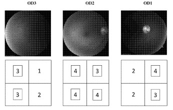
Synopsis
Patent US 9,905,008 B2 describes an automated fundus image field detection and quality assessment system. This invention addresses a critical need in ophthalmic imaging by providing a method to automatically identify the specific field of view within fundus images and evaluate their quality. This is particularly valuable for improving efficiency and diagnostic accuracy in telemedicine and large-scale retinal screening programs.
A novel aspect of this invention is its ability to automatically determine if a fundus image corresponds to a disc-centered, macula-centered, or temporal-to-macula field, or if it is an external image. This is achieved by segmenting retinal blood vessels and analyzing their morphology and position relative to the optic disc and macula. Furthermore, the system includes a comprehensive image quality assessment component that evaluates factors such as focus, illumination, and artifacts, assigning a quality score to each image. This automated assessment streamlines the process of evaluating image usability for diagnosis and follow-up.
The commercial potential for this automated system is substantial, particularly in the growing field of telehealth and ophthalmic diagnostics:
Telemedicine and Remote Diagnosis: This invention can significantly improve the workflow and reliability of tele-ophthalmology programs. By automatically identifying the correct retinal field and assessing image quality, it reduces the need for manual review and ensures that only high-quality, relevant images are used for remote diagnosis of conditions like diabetic retinopathy, glaucoma, and macular degeneration. This can lead to earlier detection and treatment, especially in underserved areas.
Mass Screening Programs: For large-scale eye screening initiatives, the ability to rapidly and accurately process a high volume of fundus images is crucial. This system can automate the initial review, flagging images that require re-imaging or immediate attention from an ophthalmologist, thereby optimizing resource allocation and reducing the burden on clinical staff.
Ophthalmology Clinics and Practices: Even in traditional clinical settings, this technology can enhance efficiency by ensuring that images captured by technicians are of sufficient diagnostic quality before the patient leaves. This minimizes the need for repeat visits and improves overall patient experience.
Clinical Research and Drug Development: In clinical trials for ophthalmic diseases, consistent and high-quality imaging is paramount for monitoring disease progression and treatment efficacy. This system can ensure standardized image acquisition and quality control across multiple sites, improving the robustness of research data.
Artificial Intelligence and Machine Learning in Ophthalmology: The automated field detection and quality assessment provide valuable pre-processing steps for AI algorithms designed to diagnose retinal diseases. By ensuring that AI models are trained and operate on properly categorized and high-quality images, the accuracy and reliability of these diagnostic tools can be greatly enhanced.
This invention offers a robust, automated solution for fundamental challenges in ophthalmic imaging, paving the way for more efficient, accurate, and scalable eye care services globally.