Home Page


Welcome to the Biomechanical Imaging Laboratory in the department of Biomedical Engineering at the Rochester Institute of Technology.
Our research is focused on the study of soft tissue biomechanics using medical imaging systems, as it relates to a variety of pathologies.
Research
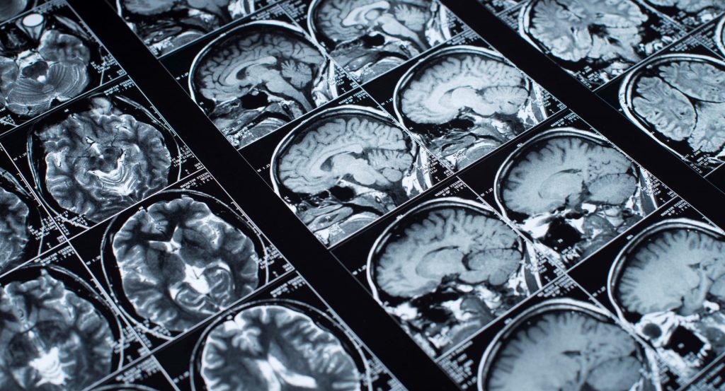
General Methods for Elastic Inverse Problems
Biomechanical imaging (BMI) is a technique that expands upon the medical imaging practice of elastography to solve the inverse problem in solid mechanics.
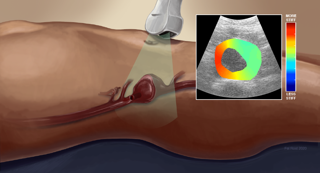
Risk Assessment of Abdominal Aortic Aneurysm
Biomechanical imaging techniques can help us measure estimates of the material property changes within aortic tissue and potentially stresses within the wall.
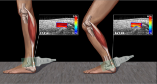
Ultrasound Elastography of Achilles Tendinopathy
Our techniques utilize common elastographic techniques such as quasi-static, quantitative elastography, measurement of large strains and shear wave imaging of tendons.
Lab News
- September 2020
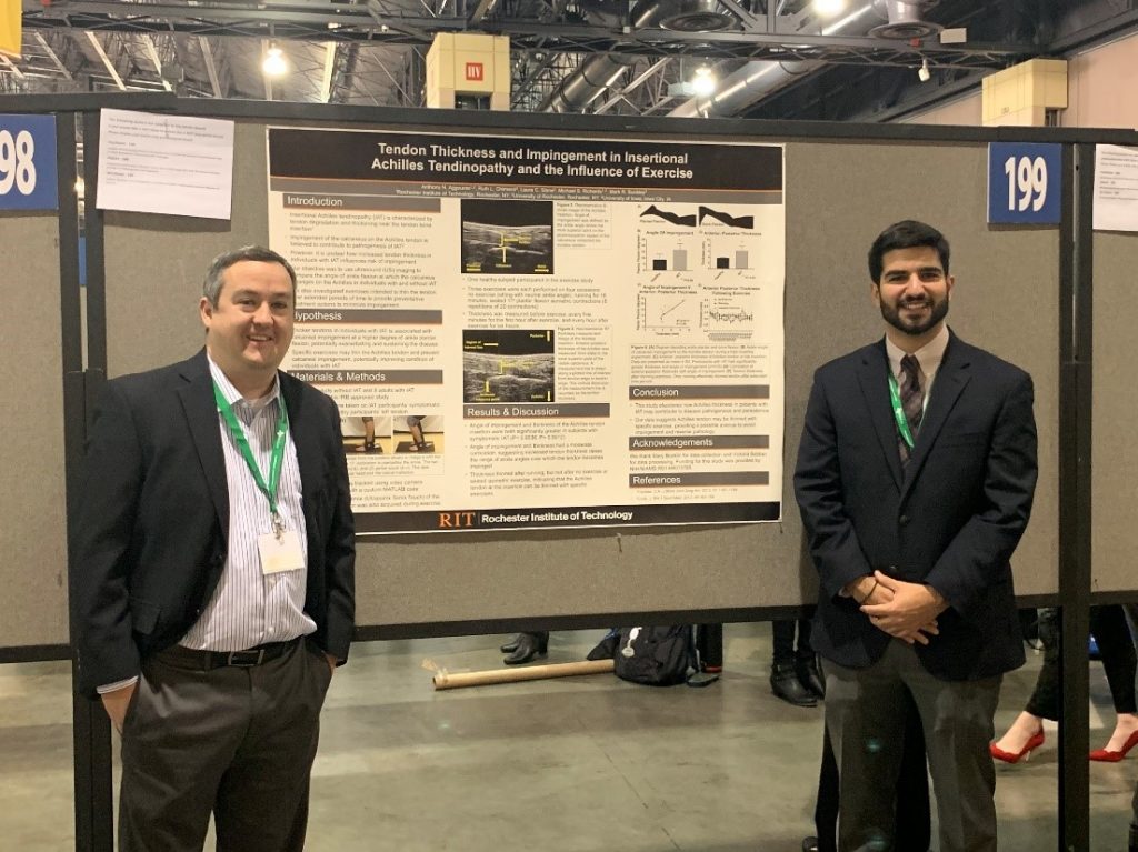
Anthony Aggouras presents his research at the annual Biomedical Engineering Society (BMES)
Tendon Thickness and Impingement in Insertional Achilles Tendinopathy and the Influence of Exercise.
- August 2020
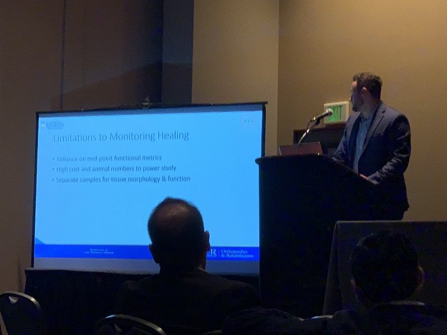
Marlin Myers presents his research at the annual Biomedical Engineering Society (BMES)
Development of Longitudinal, Non-invasive Ultrasonography to Measure Tensile Mechanical Properties in Tendon Healing.