RIT researchers receive NSF award to develop new diagnostic tool for cardiac disease
Non-invasive method increases likelihood of correctly identifying areas of deteriorating heart function
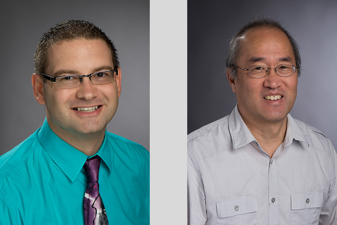
Cristian Linte, left, associate professor of biomedical engineering in RIT’s Kate Gleason College of Engineering, and Niels Otani, associate professor in RIT’s School of Mathematics.
Researchers at Rochester Institute of Technology are providing a better map to the human heart. They are developing a critical tool that will help clinicians identify damaged areas in the heart to more accurately diagnose cardiac disease.
Much like a GPS guidance system, the mapping tool may decrease the need for highly invasive, open-heart surgeries, or provide more detailed information about heart damage prior to other risky procedures.
Clinicians must first identify regions of the heart muscle that exhibit reduced contractions. Cardiac contractile activity cannot be directly measured, and physicians estimate this by measuring surrogate indicators such as diminished blood supply to certain regions or abnormal contraction and movement of heart walls, said Cristian Linte, associate professor of biomedical engineering in RIT’s Kate Gleason College of Engineering and project lead.
“Our goal is to enable non-invasive appraisal and visualization of active stresses developed in the myocardium to serve as a direct means to assess the biomechanical function of the heart,” Linte explained. He is leading a multi-disciplinary team that includes Niels Otani, associate professor in RIT’s School of Mathematics, and Suzanne Shontz, associate professor of electrical engineering and computer science at the University of Kansas.
The group received more than $850,000 from the National Science Foundation for the collaborative research on the development and validation of “A computational framework for reconstructing and visualizing myocardial active stress.” The three-year project will address a currently unexplored niche in the cardiac modeling field, specifically the reconstruction and visualization of the cardiac biomechanical activity in the form of myocardial active stresses, to enable direct appraisal of cardiac function. The team will develop a computational framework and software for cardiac biomechanical simulations using data from high-resolution cardiac MRI images. New computational algorithms developed from this data will reconstruct active stress distribution from cardiac deformations.
Together, the group proposes to quantify contraction power of the heart. They’ll estimate the stresses developed within the heart muscle by combining medical imaging and mechanical modeling of the heart. This process can help detect and localize regions that show diminished contractile activity and could become the foundation for improved diagnosis of cardiac disease.
A human heart experiences movement in response to contraction activity, but despite all areas experiencing some extent of motion, some regions don’t actually contribute as extensively to the contraction activity as nearby regions, Linte explained.
“Our goal is to dig deeper, beyond the motion alone and into the underlying stresses originating inside the tissue,” he said. “Actively contracting regions inside the heart wall generate active stresses that produce active wall motion. Non-contracting, or abnormally contracting regions, on the other hand, experience no active stress, but still experience passive motion. Our proposed technique will help identify such regions that don’t actively contract, yet still passively move, as they are dragged around by their actively contracting, actively moving neighboring regions.”
A normal heart contracts and pushes blood from the left ventricle into the aorta and further into the rest of the body. Due to various diseases, contractile capabilities are affected in certain regions of the cardiac muscle and compromise the overall heart function.
“We are developing a novel computational framework for cardiac biomechanics to reconstruct the active stresses from cardiac deformations by integrating techniques from medical image computing, high order meshing and inverse-problem biomechanical modeling,” said Linte, who has expertise in medical imaging and image computing for computer-assisted diagnosis and therapy applications. Shontz and Otani provide research expertise in high-order meshing, scientific computing and inverse problems, respectively, which are all necessary to build the biomechanical models that will become the complex, computer-aided diagnosis system that will allow clinicians to non-invasively assess the viability of the heart tissue and detect regions that experience sub-optimal contracting functions.
In addition, Linte also received $1.7 million from the National Institute for General Medical Sciences, at the National Institutes of Health, to develop and validate “Biomedical Computing and Visualization Tools for Computer-integrated Diagnostic and Therapeutic Data Science.” This five-year grant covers the wider spectrum of research being conducted in Linte’s lab specific to medical image computing, visualization algorithms and new paradigms for biomedical modeling and simulation. System validation of the computational infrastructure will be conducted through established collaborations with cardiologists at the University of Rochester Medical Center and Mayo Clinic.